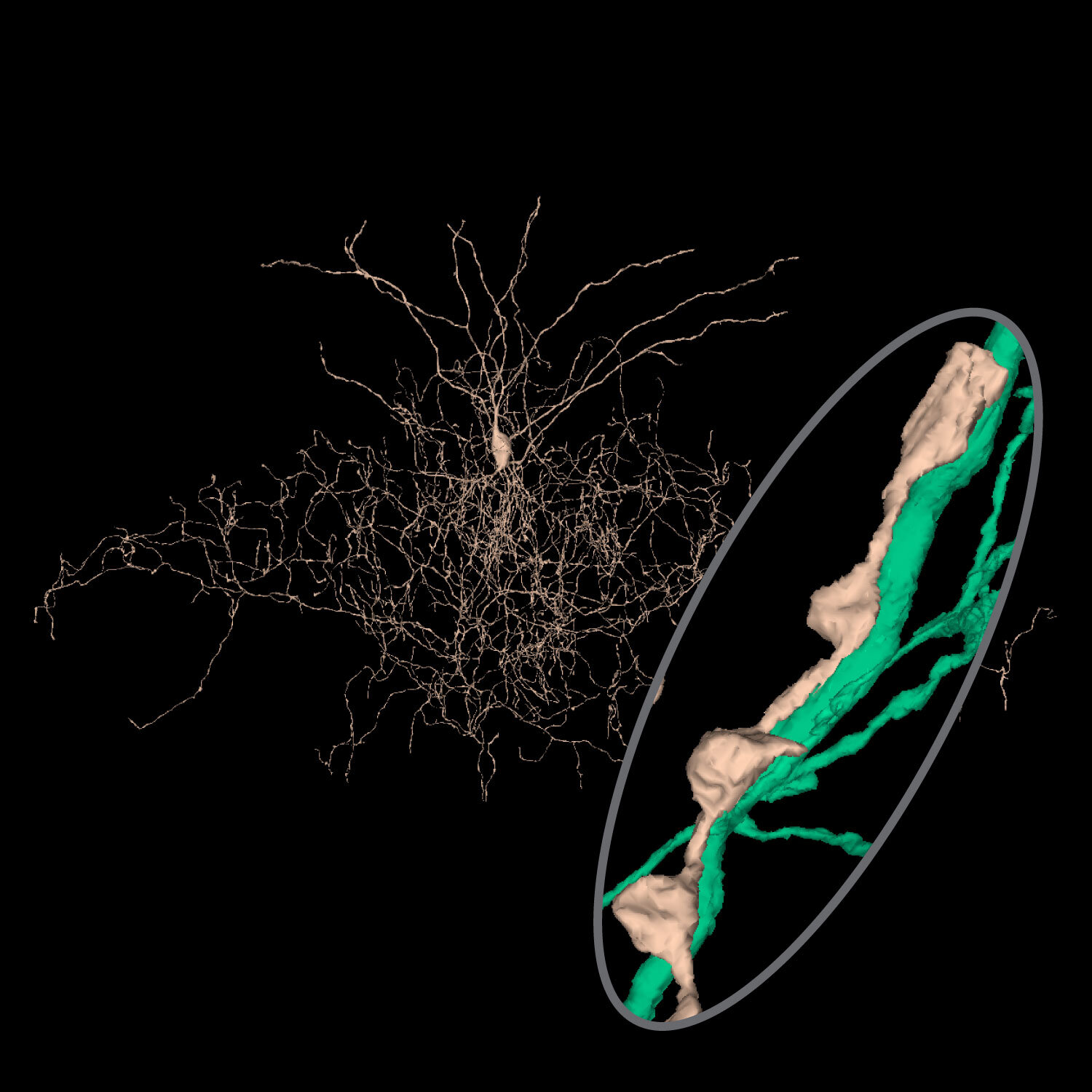
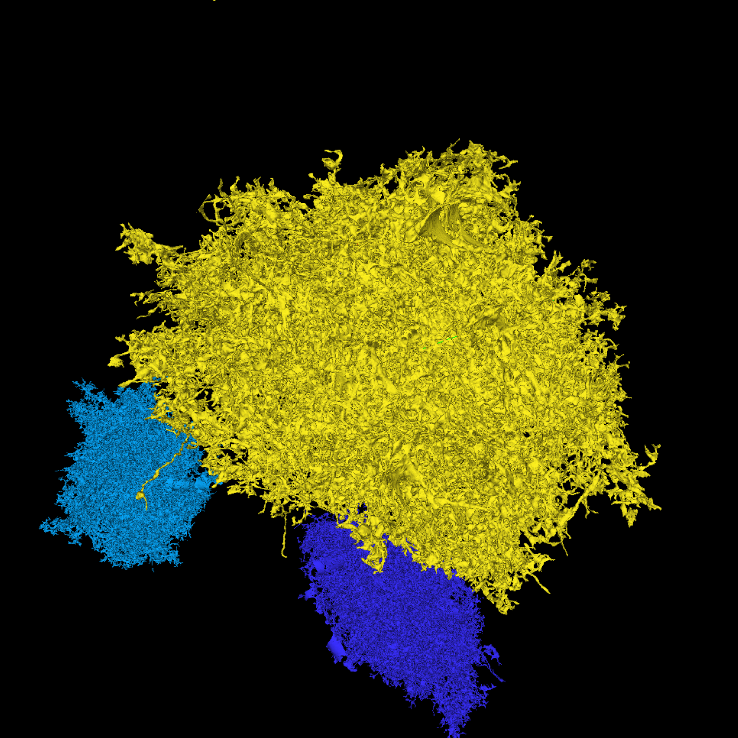
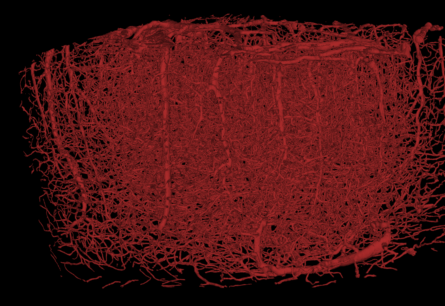
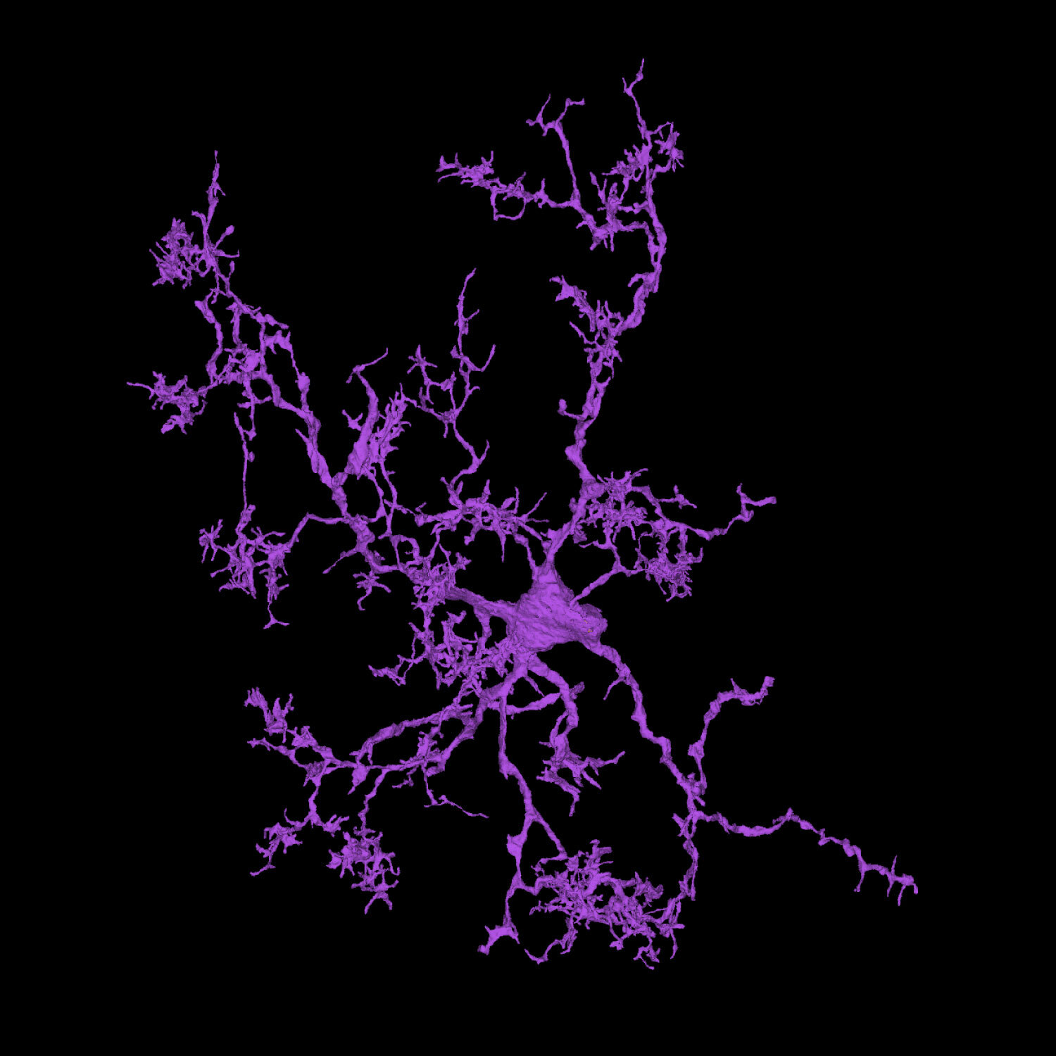
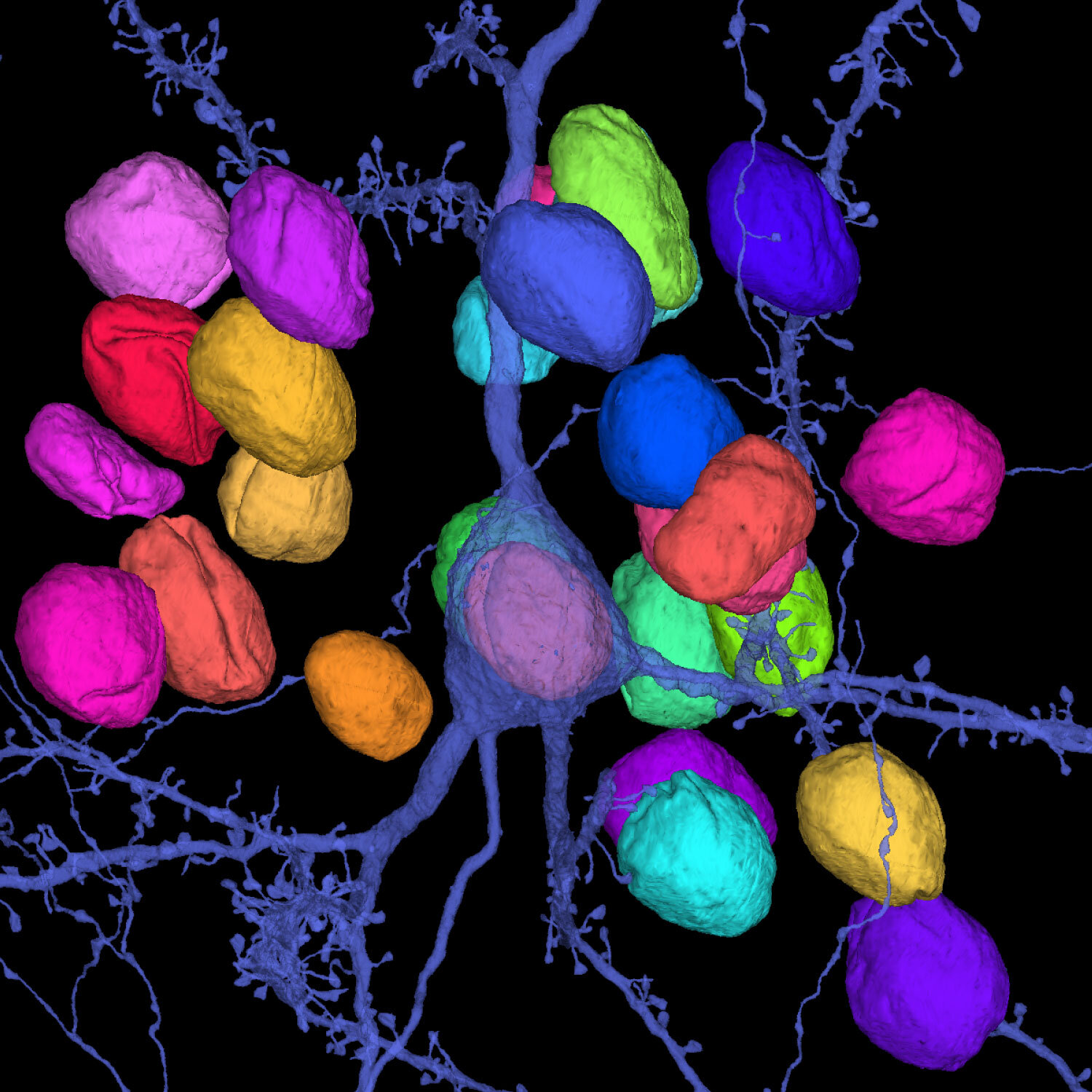
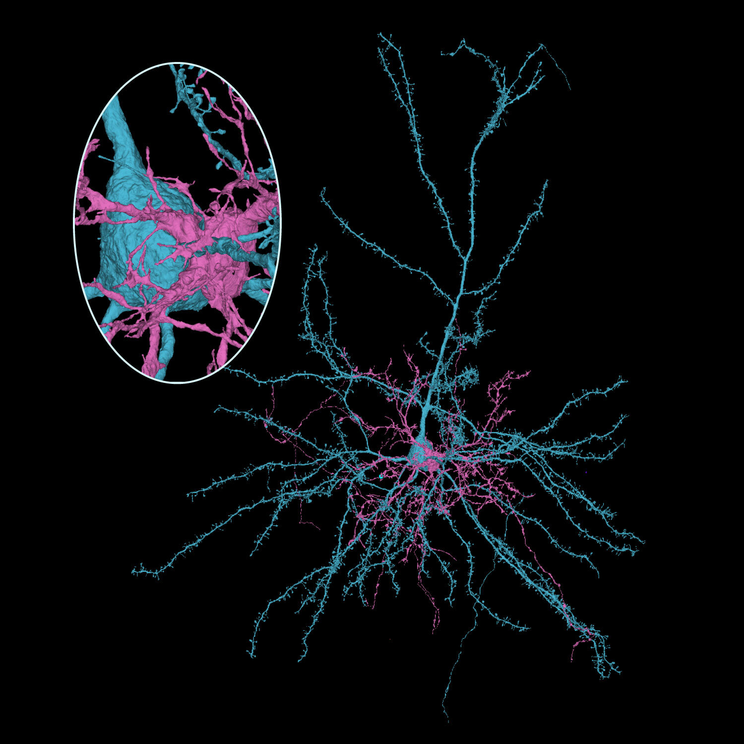
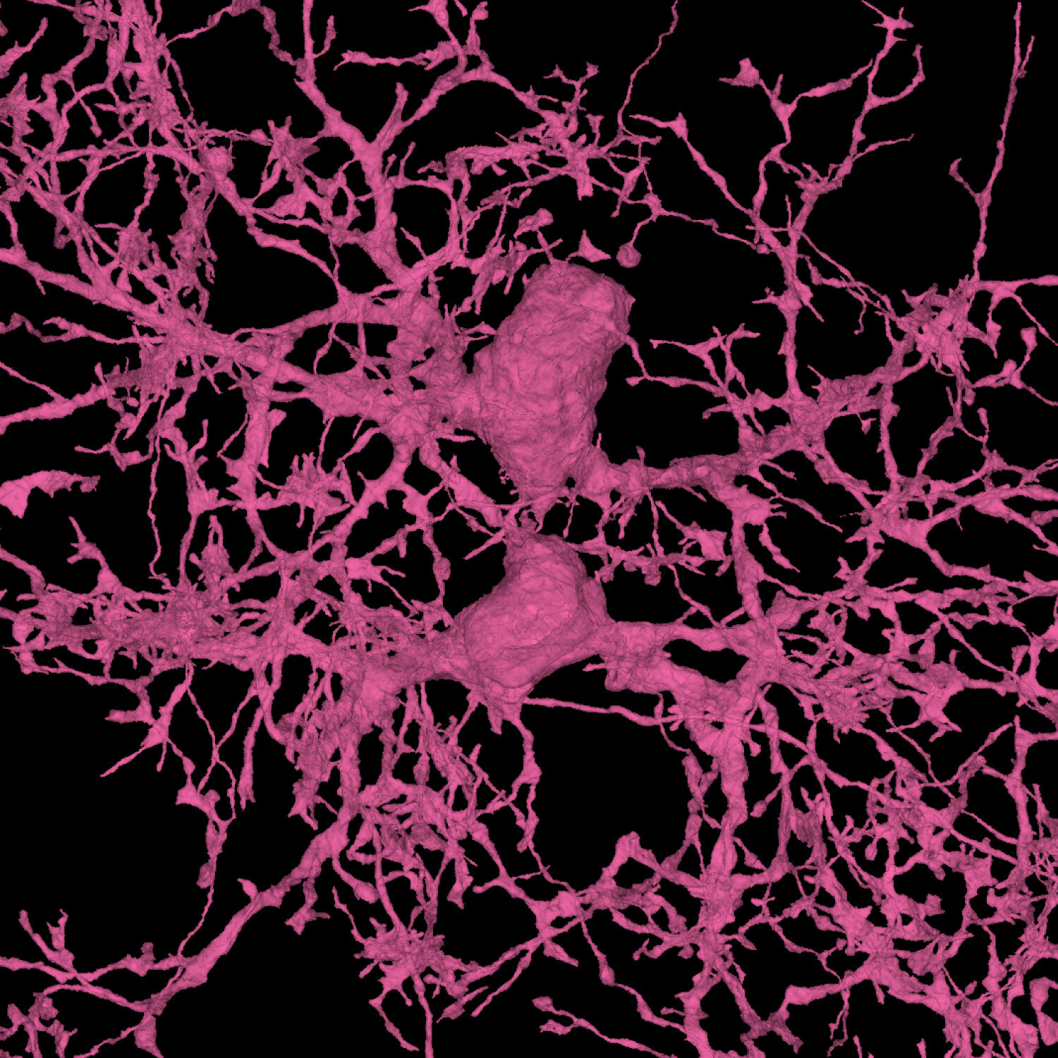
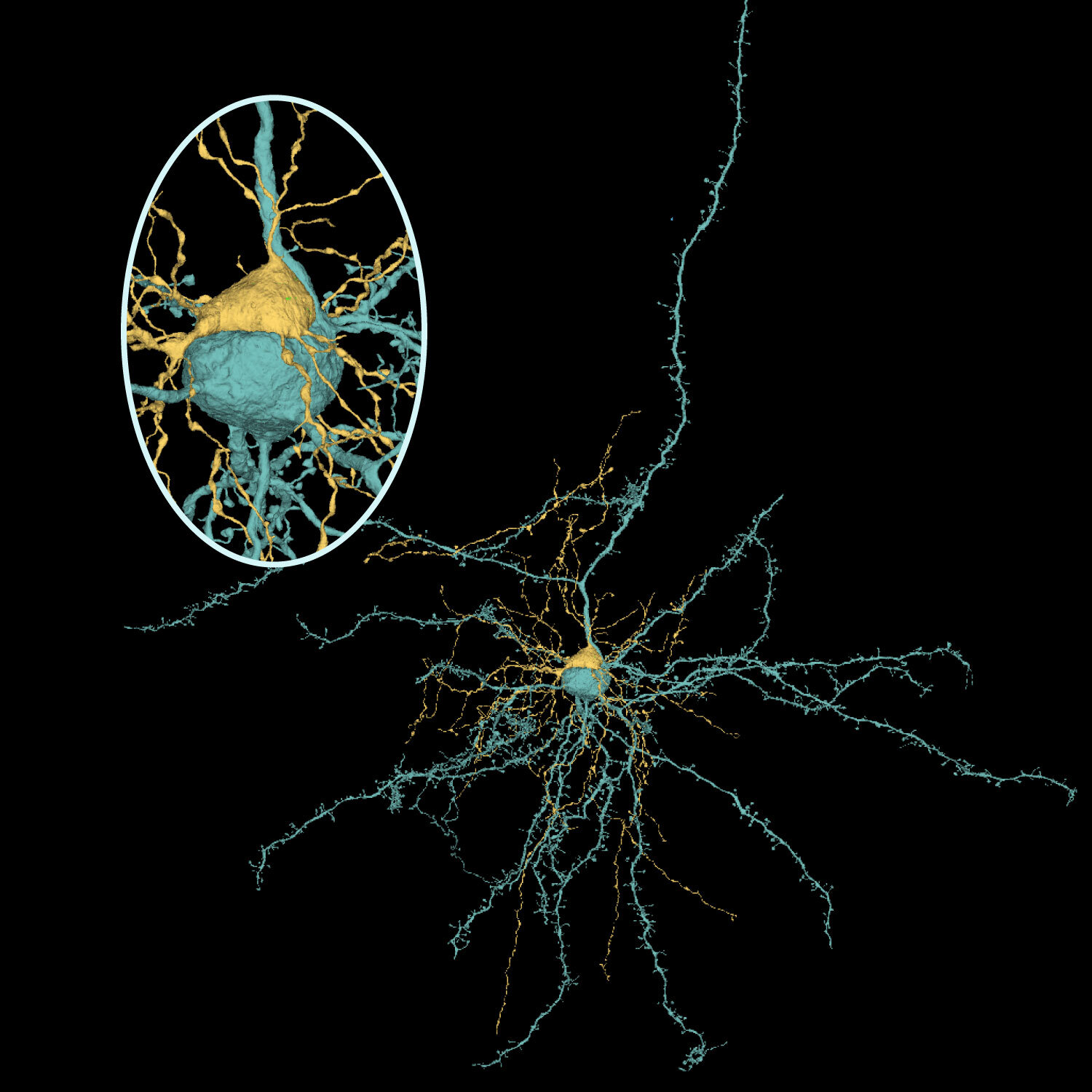
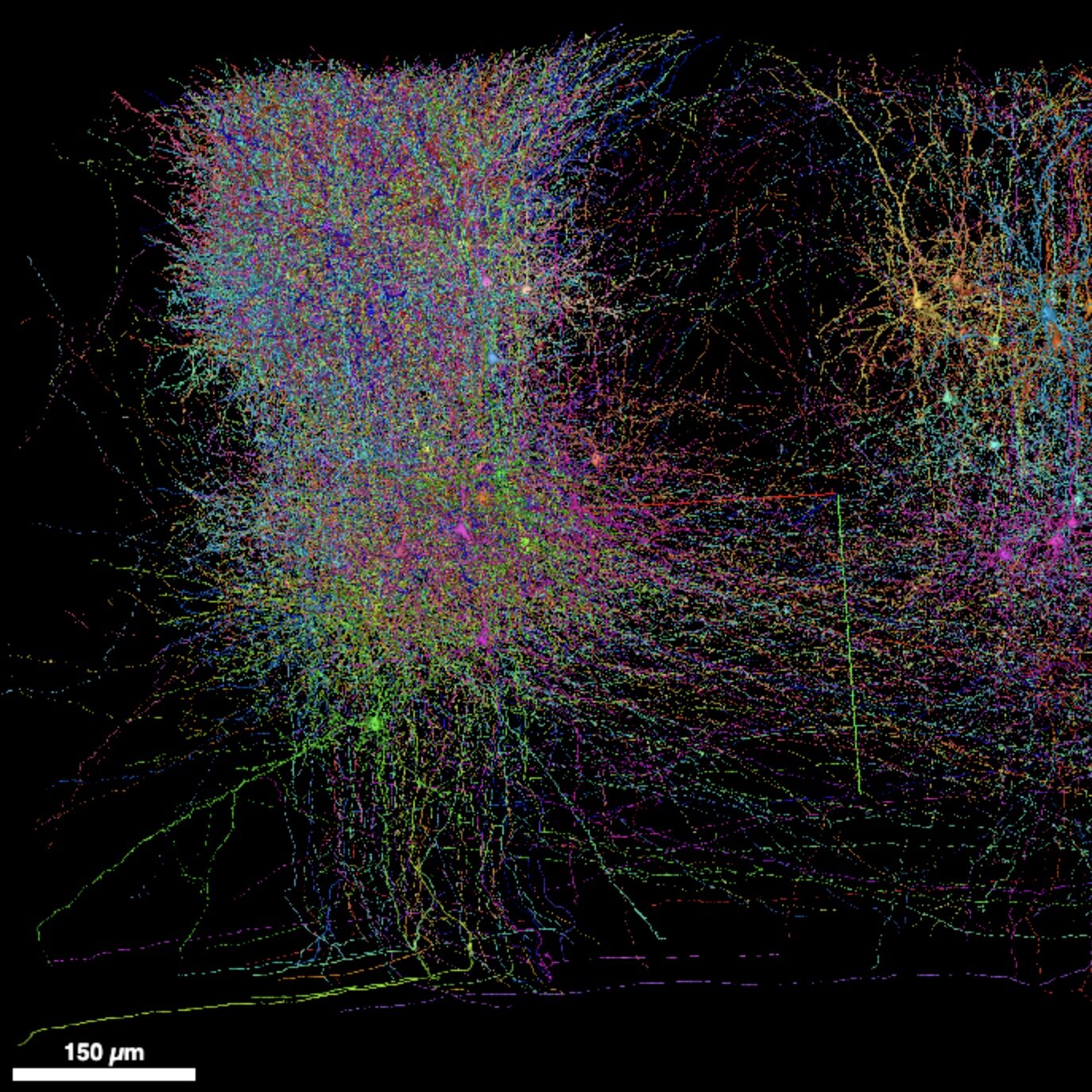
Click the images below to explore selected views in neuroglancer. Layer 5 Thick Tufted Neuron Layer 4 Cells Bipolar Cells Layer 5 NP (Near Projecting) Cell Layer 2/3 Cells Chandelier Cell with post-synaptic partners Astrocytes Basket Cells Vasculature Martinotti Cells Microglia OPC (Oligodendrocyte Precursor Cells) Synaptic Visualization Holy Soma Batman! Nuclei Forbidden Cinnamon Roll Satellite OPC hugging a Pyramidal Cell Phagolysosome in microglial branch Phagolysosome with vesicles in OPC Oligo wrapping axon Recently divided OPC Satellite Oligodendrocyte 200 cells matched to functional data. Meshes will take a while to load. Astrocyte and blood vessel Four astrocytes and vasculature. Meshes may take a while to load. Cortical columns. Meshes will take a while to load. Cell with many spines Rendered Images A rendering of a layer 5 martinotti cell with all its synaptic outputs represented as lights. This cell was reconstructed as part of a study analyzing the connectivity patterns of distinct types of layer 5 martinotti types. A rendering of a layer 5 martinotti cell with all its synaptic outputs represented as lights. This cell was reconstructed as part of a study analyzing the connectivity patterns of distinct types of layer 5 martinotti types. A rendering of 4 layer 5 thick tufted pyramidal neurons. The connectivity of these cells was explored in the linked paper. A rendering of layer 5 thick tufted pyramidal neurons. A rendering of layer 5 thick tufted pyramidal neurons. A zoomed in view of neural processes of 4 distinctly colored neurons. A rendering of layer 5 thick tufted pyramidal neurons where a subset of synapses are light up like lights on a christmas tree. Still frame from a movie that won an award in the BRAIN Initiative's Show Us Your Brains contest. A close up rendering of an oligodendrocyte precursor cell enveloping the synaptic contact between an axon and a dendrite. The role of these cells in consuming synapses was first described based on observations in this dataset. The paper is linked. A rendering of the dataset including thousands of individual neurons, but still only showing a small subset of the cells that are in the dataset. The gray portion shows an electron micrograph of a single section at the back of the dataset. The somatic region of a subset of cells that were part of a study about the patterns of inhibitory connectivity onto excitatory cells in cortex are rendered in different colors according to their cell types. The gray backing shows the electron micrograph of the larger dataset. The paper is linked for more information. A rendering of several cells in the dataset that is used as the background for the main page on the cubic millimeter dataset. A rendering of a chandelier cell with its output boutons lit up as lights. The connectivity of this cell type was examined in the linked paper. A rendering of a chandelier cell with its output boutons lit up as lights. The connectivity of this cell type was examined in the linked paper. A rendering of a segment of dendrite as if it were made of glass. A simple rendering of a layer 5 thick tufted cell. A rendering of the blood vessels in the brain. The dataset has created opportunities for studying the relationship between the vasculature and the brain as highlighted in the linked publication.